


Perihal Interactive CT & MRI Anat.Lite
★Lite version★
This is the free Lite version of "Interactive CT and MRI Anatomy".
The function is restricted.
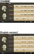
You can only see the transverse CT images of the head.
Please check the operation before purchasing the full version.
★ Details ★
This application is developed for medical students, interns, residents, doctors, nurses, and radiology technicians to understand the essential anatomical terms of the body.
You can learn anatomy by answering the terms by step-to-step questions using a total of 241 CT and MRI images.
A total of 17 images of 3D-CT, MRA and plain X-ray film(particularly the extremities) are included as references.
Other reference images include coronary artery segments defined by the American Heart Association(AHA), pulmonary segments, and liver segments(according to Couinaud classification).
You can enlarge all the images by simple manipulation.
★ Major functions ★
There are 4 major functions.
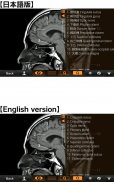
-1) Anatomical mode
Anatomical terms are overlaid on the images.
It can be used as the anatomical atlas.
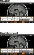
-2) Quiz mode type 1
You select the part of the image by using anatomical term.
Questions will basically appear randomly.
-3) Quiz mode type 2
You select the anatomical term by the part of the image.
Questions will basically appear randomly.
-4) Index
You can find the specific images by using anatomical terms.
★ Intended users ★
-Medical students
-Interns and residents
-Doctrors
-Nurses
-Radiology technicians
-All those who are intrested in CT and MRI anatomy
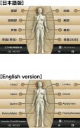
★ Images(a total of 258 images) ★
Images basically include horizontal, coronal, and sagital planes.
-Head(36 images including CTA and 3D-CT)
-Neck(24 images)
-Spine(19 images including plain X-ray films)
-Chest(61 images including 3D-CT images)
-Abdomen (37 images)
-Pelves: male (9 images)
-Pelvis: female (11 images)
-Extremities (shoulder, hand, elbow, hip joint, knee, foot) (61 images including plain X-ray films)
Editors
Toshiaki Nitori, M.D. (Professor of Radiology, Kyorin University, School of Medicine)
Yasuo Sasaki, M.D. (Manager of diagnostic radiology, Iwate Prefectural Central Hospital)
</div> <div jsname="WJz9Hc" style="display:none">★ versi Lite ★
Ini adalah versi Lite bebas "CT Interaktif dan MRI Anatomi".
Majlis tersebut adalah terhad.
Anda hanya boleh melihat melintang CT imej kepala.
Sila semak operasi sebelum membeli versi penuh.
★ ★ Butiran
Aplikasi ini dibangunkan untuk perubatan pelajar, pelatih, penduduk, doktor, jururawat, juruteknik dan radiologi untuk memahami terma anatomi yang penting dalam badan.
Anda boleh belajar anatomi dengan menjawab terma-terma dengan langkah-langkah untuk soalan menggunakan sejumlah 241 CT dan MRI imej.
Seramai 17 imej 3D-CT, MRA dan jelas X-ray filem (terutama kaki) disertakan sebagai rujukan.
Rujukan imej lain termasuk segmen arteri koronari yang ditakrifkan oleh Persatuan Jantung Amerika (AHA), segmen paru-paru, hati dan segmen (mengikut klasifikasi Couinaud).
Anda boleh membesarkan semua imej dengan manipulasi mudah.
★ ★ fungsi utama
Terdapat 4 fungsi utama.
-1) Mod anatomi
Segi anatomi yang bertindan pada imej-imej.
Ia boleh digunakan sebagai atlas anatomi.
-2) Kuiz mod jenis 1
Anda memilih sebahagian daripada imej dengan menggunakan istilah anatomi.
Soalan pada dasarnya akan muncul secara rawak.
-3) Kuiz mod jenis 2
Anda pilih istilah anatomi oleh sebahagian daripada imej.
Soalan pada dasarnya akan muncul secara rawak.
-4) Indeks
Anda boleh mencari imej-imej tertentu dengan menggunakan istilah anatomi.
Pengguna Dimaksudkan ★ ★
Pelajar -Medical
-Interns Dan penduduk
-Doctrors
-Nurses
Juruteknik -Radiology
-Semua Mereka yang intrested dalam CT dan MRI anatomi
★ Imej (sejumlah 258 imej) ★
Imej pada dasarnya termasuk kapal terbang mendatar, korona, dan sagital.
-Head (36 gambar termasuk CTA dan 3D-CT)
-Neck (24 gambar)
-Spine (19 gambar termasuk dataran filem X-ray)
-Chest (61 gambar termasuk imej 3D-CT)
-Abdomen (37 gambar)
-Pelves: Lelaki (9 gambar)
-Pelvis: Perempuan (11 gambar)
-Extremities (Bahu, tangan, siku, sendi pinggul, lutut, kaki) (61 gambar termasuk dataran filem X-ray)
Editor
Toshiaki Nitori, MD (Profesor Radiologi, Kyorin University, Sekolah Perubatan)
Yasuo Sasaki, MD (Pengurus radiologi diagnostik, Iwate Hospital Wilayah Tengah)</div> <div class="show-more-end">
Interactive CT & MRI Anat.Lite - Versi 1.2
(04-07-2015)Interactive CT & MRI Anat.Lite - Maklumat APK
Versi APK: 1.2Pakej: com.libroscience.www.ctmri.lightAplikasi dalam kategori sama
Anda juga mungkin suka...


